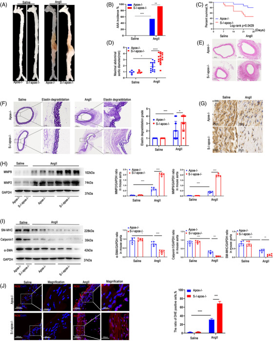
FIGURE 2.

Knockout (KO) of SDC4 promoted AAA formation under Ang II stimulation. All mice were administered AngII or saline for 28 days. (A) Representative images showing the macroscopic features of AngII‐induced AAAs. (B and C) The AAA incidence (B) and survival curve (C) of Ang II‐induced apoe‐/‐ mice (n = 15) compared with SDC4‐/‐apoe‐/‐ mice (n = 16). Saline infusion did not induce AAA formation (n = 10). (D) Statistical analysis of the maximal abdominal aortic diameter in Ang II‐ and saline‐infused mice. (E and F) Representative photographs of H&E staining (E), elastica van Gieson (EVG) staining and elastin degradation scores (F) in abdominal aortas from AngII‐ and saline‐infused mice (n = 5) (scale bars, 300 μm and 100 μm) (magnified photographs). (G) Immunohistochemical analysis of α‐SMA‐positive cells in SDC4‐/‐apoe‐/‐ and apoe‐/‐ mice after AngII or saline treatment (scale bars, 25 μm). (H) Representative western blots of MMP9 and MMP2 and densitometric analysis of AngII‐ and saline‐infused mice (n = 5). (I) Representative western blots of α‐SMA, Calponin1 and SM‐MHC and densitometric analysis of saline‐ and AngII‐infused mice (n = 5). (J) The level of reactive oxygen species (ROS) in the abdominal aortas of AngII‐ and saline‐infused mice was evaluated by dihydroethidium (DHE) staining and quantified by determining the ratio of DHE‐positive cells (n = 5) (scale bars, 500 μm and 100 μm) (magnified photographs)
