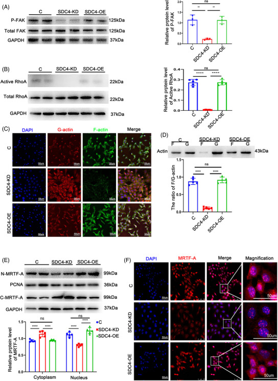
FIGURE 5.

SDC4 was crucial in maintaining the contractile phenotype of vascular smooth muscle cells (VSMCs) via the RhoA‐MRTF‐A axis. (A) Representative western blots of P‐FAK and densitometric analysis of control, SDC4‐KD and SDC4‐OE cells (n = 5). (B) Representative western blots of active RhoA and densitometric analysis of control, SDC4‐KD and SDC4‐OE cells (n = 5). (C) Representative phalloidin staining of G‐actin and F‐actin in control, SDC4‐KD and SDC4‐OE cells (n = 5) (scale bars, 50 μm). (D) Relative protein expression of G‐actin and F‐actin was determined by western blot analysis with a commercial kit (cytoskeleton, Cat #BK037) in control, SDC4‐KD and SDC4‐OE cells (n = 5). (E) Representative western blots of MRTF‐A and densitometric analysis of proteins in the cytoplasm and nucleus in control, SDC4‐KD and SDC4‐OE cells (n = 5). (F) Representative immunofluorescence images of nuclear MRTF‐A in control, SDC4‐KD and SDC4‐OE cells were analysed by confocal microscopy (n = 5, scale bars, 50 μm)
