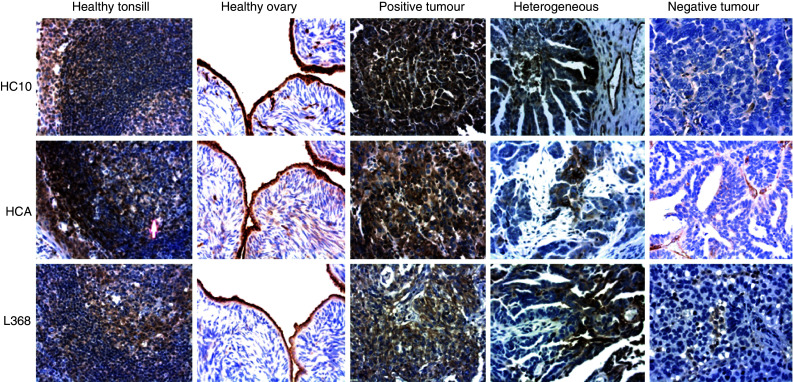
Fig. 1.

Representative examples of immunohistochemical staining patterns of different MHC components in healthy tissues using the mAbs HC-10 and HCA detecting the HLA class I HC and mAb L368 detecting β2-m (reading the figure horizontally). Vertically and from left to right; healthy tonsil and ovary, tumour tissue with the majority of stained cells (positive), heterogeneous and negative (clearly positive infiltrating cells in the stroma) staining. Original magnification ×400
