Fig. 1.

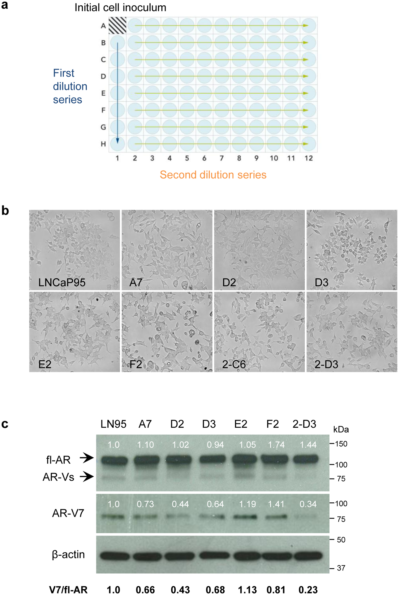
Generation of clones from the LNCaP95 parental cell line. a Schematic summarizing the serial dilution of LNCaP95 cells in a 96-well plate. Arrows represent the first and second dilution series. b Micrographs depicting the cell morphology of LNCaP95 parental cells and isolated clones. LNCaP95 cells have an epithelial cell morphology with pronounced dendritic-cell-like extensions, whereas several of the isolated clones had a unique flattened morphology. c Western blot analysis of fl-AR and AR-V7 expression in the isolated clones. Numbers above the protein bands indicate the relative fl-AR and AR-V7 expression after normalizing to β-actin. The ratio of AR-V7 expression relative to fl-AR is indicated below
