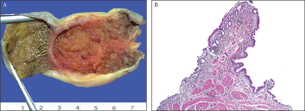
Figure 1:
(A) There are anatomic polypoid folds such as Phrigian cap deformity as well as the valves at the neck/cystic-duct region and those occur due to impacted stones, all of which were excluded from the study. Typically, these form a pencil-like projection that come out of the surface with an angle. (B) Microscopically, these tend to have a well defined and organized muscle core at its center that tapers towards the tip, and a normal mucosal covering that is almost equidistant from the muscle similar to the mucosa elsewhere.

