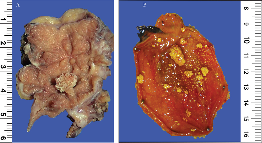
Figure 2:
(A) A 0.7 cm fibromyoglandular polyp is seen in the background of a polypoid/nodular gallbladder mucosa with prominent wall thickening and gallstones (See Figure 3 for microscopy).
(B) A case of multiple cholesterol polyps, which were typically seen in gallbladders devoid of any significant chronic changes (notice the non-polypoid mucosa). Because of the presence of cholesterol-laden macrophages, which were often in abundance, they had the distinctive yellow color in macroscopic examination. They were characterized by a striking cauliflower pattern which was highly consistent in virtually all examples.

