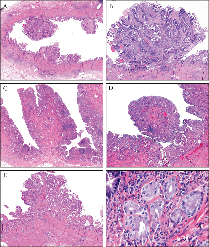
Figure 3:
Fibromyoglandular polyps. (A) These occurred in the background of mucosal injury and were commonly multifocal. They had a spectrum of glandular and stromal components with variable muscle participation. The glands were also distributed variably. (B) Some had more fibrotic stroma and relatively evenly distriuted glands. (C) Others had substantial amount of muscular stroma with mucosally-oriented glandular components. (D) Inflammation can be prominent in some. (E) Lobulated collections of glands could be seen and (F) these were often of pyloric gland type.
[Microscopic photographs of Figure 2a]

