. 2021 Jan 6;11(4):1939–1951. doi: 10.1039/d0ra09992a

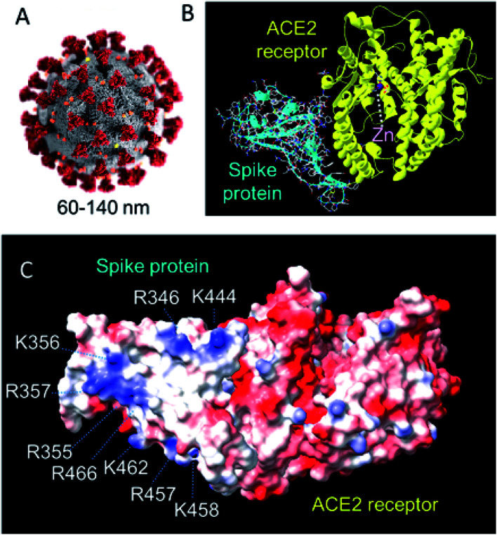
Fig. 12. (A) Structure of SARS-CoV-2. The virus has a diameter of ca. 60–140 nm with 9–12 nm crown-like spikes of the transmembrane spike (S) glycoprotein on its surface.64 This S protein forms trimers and facilitates binding to angiotensin-converting 2 (ACE2), a zinc enzyme on host respiratory tract cells.65–68 Reproduced with permission from CDC/Alissa Eckert, MSMI; Dan Higgins, MAMS.69 (B) The 2.5 Å resolution X-ray crystal structure of coronavirus spike receptor-binding domain (largely β-sheet) complexed with its receptor ACE2 (largely α-helical, based on pdb 6lzg, courtesy of Claudia Blindauer). ACE2 is a protease. The catalytic active-site Zn(ii) (coordinated to 2His, Glu and H2O) is labelled. 13 polar residues are involved in hydrogen bonds and/or salt bridges with the SARS-CoV-2. (C) Surface electrostatic charge distribution in the structure shown on B (red, −ve; blue, +ve). The glycan chains are not defined in this structure. Notable is the cluster of positively-charged Lys and Arg residues on this face of the spike protein that might be a target for recognition by e.g. negatively charged POMs. (pdb 6lzg, prepared using UCSF ChimeraX).

Fig. 12