FIG 4.
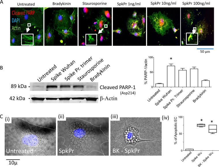
SARS-CoV-2 spike protein (SpkPr) and PARP-1 cleavage in endothelial cells. (A) Representative SR-FLICA (red), actin, and DAPI (blue) composite images of individual untreated, staurosporin (1 μM positive control), and SpkPr (1, 10, and 100 ng/ml) treated endothelial cells for 72 h (Inset: actin disassembly). (B) Representative Western blot data showing cleaved PARP-1(asp214) after 72 h of exposure of recombinant spike protein-Wuhan, & recombinant spike Pr. Active trimer (100 ng/ml, n = 3). (C-i–iv), Representative DIC and DAPI (blue) composite images of individual EC following indicated treatments. Percentage of morphologically apoptotic ECs after indicated treatments. (n > 100 cells, NS, not significant; *, P < 0.05).

