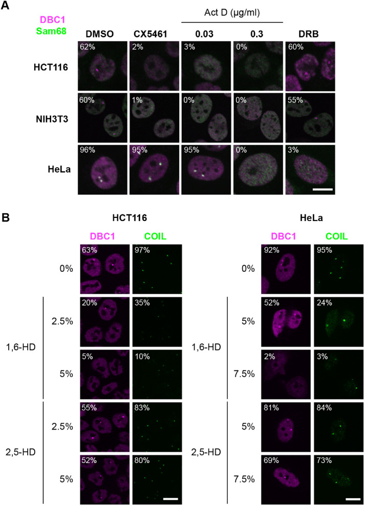
FIGURE 1:
Features of the DNB. (A) DNB formation requires RNAPI transcription. IF analysis of both DBC1 (magenta) and Sam68 (green) was performed in either HCT116, NIH3T3, or HeLa cells treated with CX5461 (2 μM), Act D (0.03 and 0.3 μg/ml), or DRB (100 μM). The control cells were treated with DMSO. DNB (DBC1 signal) are detectable in HCT116 and NIH3T3 cells. SNB (DBC1 and Sam68 signals overlap) are detectable in HeLa cells. (B) IF analysis of DBC1 (magenta) and COIL (green) was performed in both HCT116 and HeLa cells treated with 0%–7.5% 1,6-HD or 2,5-HD. DNB and SNB were detected in DBC1. Cajal bodies were detected in COIL. Both nuclear body component signals dispersed upon 1,6-HD treatment. The cell populations (%) of summary NBs (DNB, SNB, and Cajal body) signals are shown in both A and B (>100 cells, n = 5). Bars, 10 μm.

