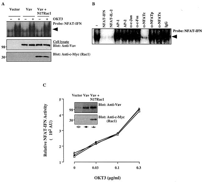
FIG. 3.
Effects of Vav on DNA-binding and transcriptional activities of NFAT-IFN. (A) Cells were transfected with empty pEF vector, Vav, or Vav plus N17Rac1. After 24 h, cells either were left unstimulated or were stimulated with cross-linked OKT3. Nuclear extracts were prepared and analyzed by an EMSA using a 32P-labeled NFAT-IFN probe (top panel). Total cellular extracts from the same groups were immunoblotted with anti-Vav (middle panel) or anti–c-Myc (bottom panel) antibodies. The results are representative of three separate experiments. The arrowhead indicates the specific NFAT-IFN complex. (B) A similar EMSA was conducted using a nuclear extract from OKT3-stimulated, Vav-transfected cells in the absence (−) or presence of the indicated unlabeled competing oligonucleotides, antibodies, or control IgG. A similar inhibition pattern was observed with an extract from unstimulated, Vav-transfected cells (data not shown). (C) Cells were cotransfected with an NFAT-IFN–Luc reporter plasmid plus empty vector, Vav, or Vav plus N17Rac1. Stimulation and reporter activity determinations were done as described in the legend to Fig. 1. AU, arbitrary units. The data shown are representative of four experiments. Samples of the same lysates were analyzed for the expression of Vav or Rac1 by immunoblotting with anti-Vav or anti-c-Myc MAbs (insets). The positions of molecular weight standards (in thousands) are shown.

