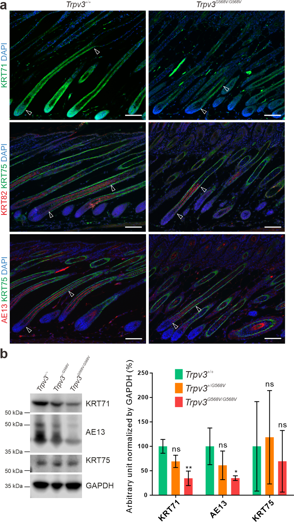
Figure 3. Impaired hair follicle differentiation in Trpv3 knock-in mice.

(a) Immunofluorescence labeling of KRT71 (green), KRT75 (green), KRT82 (red), and hair cortex keratins detected by the AE13 antibody (red) in dorsal skins of P12 Trpv3+/+ and Trpv3G568V/G568V littermates. Arrow heads point to the portions of hair follicles positive for KRT71, KRT82, and AE13, respectively. Nuclei were stained with DAPI (blue). (b) Western blotting of KRT71, hair cortex keratins (AE13), and KRT75, and quantifications. Whole length images can be found in Supplementary Figure S6. n = 3 mice per genotype. Data are presented as mean ± SD. * P < 0.05. ** P < 0.005. ns, not significant. One-way ANOVA analysis was followed by Dunnett’s Multiple Comparison Test. Scale bar = 100 μm (a).
