Fig. 1.
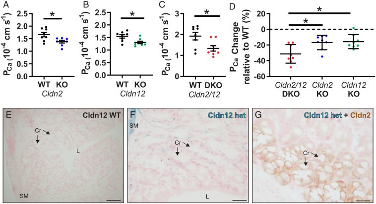
Cldn2 and Cldn12 confer independent Ca2+ permeability to the proximal colon. PCa measured ex vivo in Ussing’s chambers across the proximal colon compared to WT of Cldn2 KO (A) (n = 8 per group and P = 0.016); Cldn12 KO (B) (n = 8 per group and P = 0.012); and Cldn2/12 DKO (C) (n = 8 per group and P = 0.019). Full results of bionic dilution potential experiments on small intestine and colon of DKO mice are in SI Appendix, Fig. S2 and Tables S1–S4. Means were compared by Student’s t test. (D) Data from A–C expressed as the percentage change in PCa relative to WT for each genotype. Data are presented as mean ± SD. One-way ANOVA with Dunnett correction for multiple comparisons to compare mean from DKO mice to Cldn2 KO (P = 0.017) and Cldn12 KO (P = 0.010) was performed. The Cldn12 coding exon was replaced with a LacZ cassette. (E and F) We used this to localize Cldn12 expression by X-gal staining of colon from Cldn12 WT (E) and Cldn12 heterozygous mice (F). Staining is present in colonic crypt epithelial cells in Cldn12 heterozygous mouse (cyan). (Scale bars, 100 µm [E] and 25 µm [F]). (G) X-gal staining of colon for Cldn12 (cyan) and immunohistochemical staining for claudin-2 (brown) from a Cldn12 heterozygous mouse. Cr = crypts, SM = smooth muscle, and L = colonic lumen. (Scale bar, 25 µm.) *P < 0.05.

