FIGURE 1:
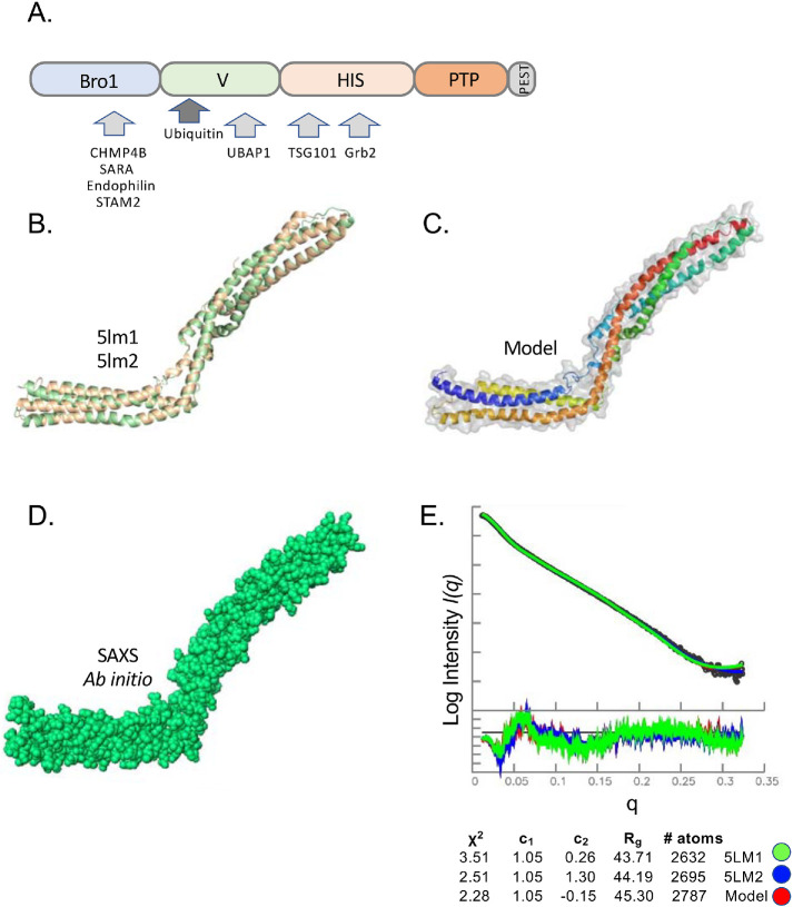
SAXS analysis of HD-PTP V domain. (A) Domain organization of HD-PTP including N-terminal Bro1 domain (residues 8–361), V domain (residues 366–704), histidine/PRR domain (residues 705–1128), phosphatase domain (residues 1192–1452), and C-terminal PEST domain (residues 1509–1636). Proteins known to interact with those domains are indicated. (B) Structure cartoon of the crystal structures of the HD-PTP V domain (PDB: 5LM1 [green], 5LM2 [wheat]). (C) Solution model for the HD-PTP V domain based on chimera of crystal structures and computational modeling missing loops colored from N- to C-termini using blue–red spectrum. (D) Ab initio envelope of the HD-PTP V domain in solution from SAXS data. (E) Guinier region of SAXS data of VHDPTP (4 mg/ml) as well as residuals to confirm little to no aggregation. At the bottom is the experimental and simulated pair distance distribution function P(r). Rg and Dmax determination from P(r) curves and χ2 values comparing experimental data with predicted P(r) curves of crystal structures and the solution model in C.

