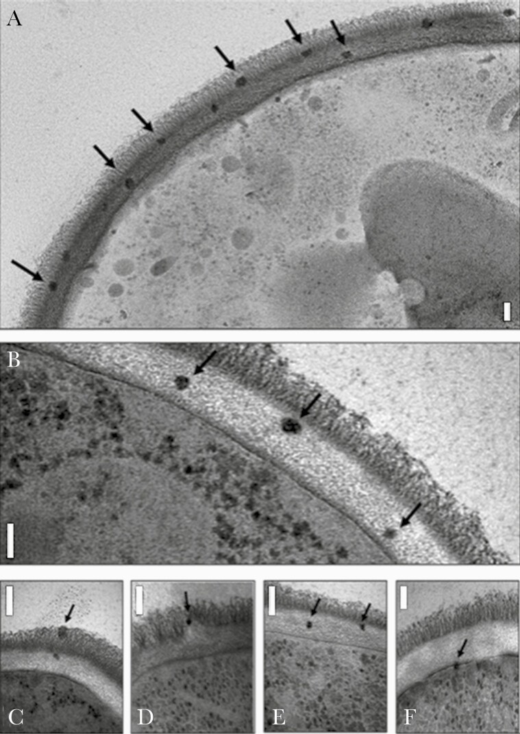
Figure 1.
Electron micrograph images of Candida albicans incubated with AmBisome, showing intact liposomes in the outer (A, C, and D) and inner (A, B, C, and E) cell wall and at the cell membrane (F), indicated by arrows. The granular particles in the cytoplasm are ribosomes, not liposomes. Bars represent 100nm. The photomicrograph originally appeared in Walker et al [19] (open access journal published by the American Society of Microbiology).

