. 2021 Dec 7;13(1):247–256. doi: 10.1039/d1sc05057e

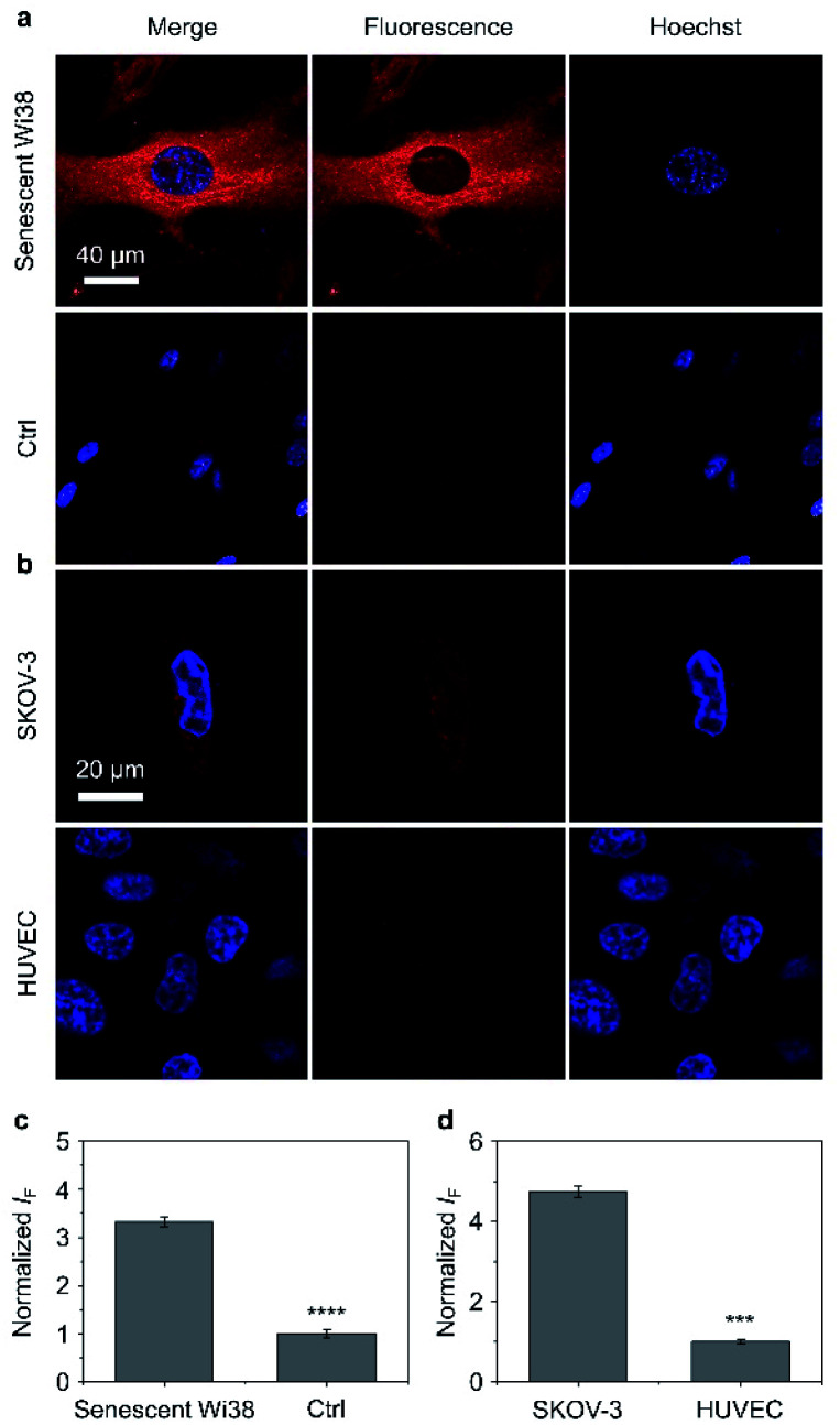
Fig. 4. (a) Fluorescence imaging and (c) quantification of senescent Wi38 cells and untreated Wi38 cells (ctrl) after incubation with TD-Gal6 (10 μM) for 40 min. (b) Fluorescence imaging and (d) quantification of SKOV-3 (human ovarian carcinoma cells) and HUVEC (human umbilical vein endothelium cells) cells after incubation with TD-Gal6 (10 μM) for 40 min. The excitation and emission channels used for TD-Gal6 are 488 nm and 580–620 nm, respectively; those for Hoechst 33342 are 405 nm and 440–480 nm, respectively. S. D. means standard deviation (n = 3). ***P < 0.001, ****P < 0.0001. Statistical analysis was performed using Student's unpaired t-test. All experiments were repeated three times with representative data shown.

Fig. 4