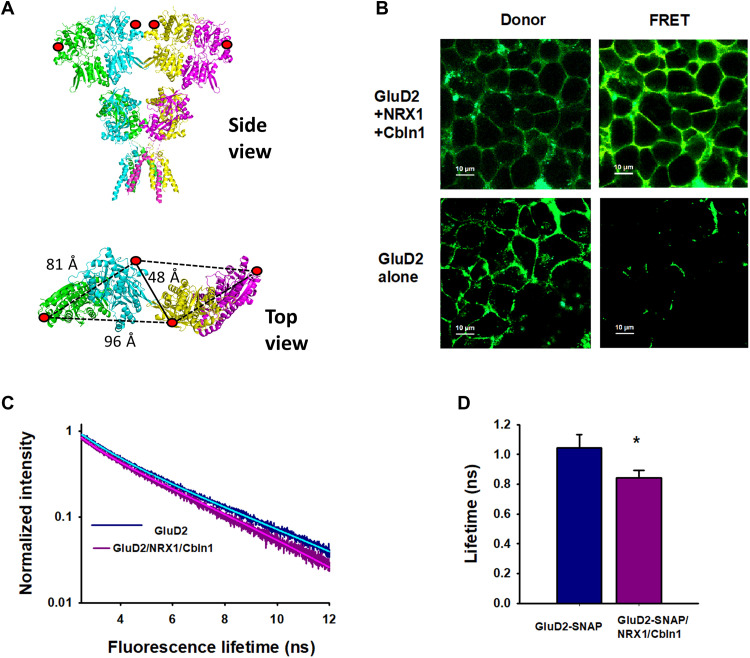
Fig. 4. Cbln1 and NRX1 induce tighter packing of the N-terminal domains of GluD2.
(A) Structure of GluD2 (modeled on GluD1; PDB: 6KSS) showing SNAP tag incorporation at position 302 (red circles) and distance between dimers. Side view (top) and close-up top-down view of 302 N-terminal domain residues (bottom). (B) Fluorescent images of HEK-293T cells transfected with GluD2 alone or GluD2, NRX1, and Cbln1 showing the intensity of donor and FRET fluorescence. (C and D) Representative fluorescence lifetimes of donor- and acceptor-labeled cells expressing GluD2 alone or GluD2, Cbln1, and NRX1; solid lines show two exponential fits. (D) Donor lifetimes from at least three different days. Data represent mean lifetimes with SEs of the mean, *P = 0.00052.

