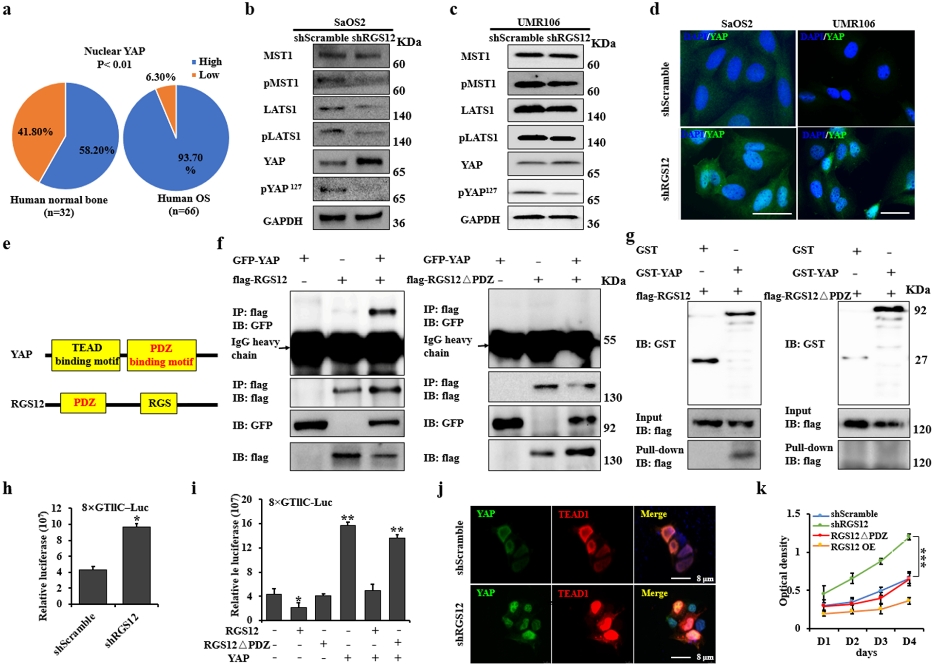
Fig. 4. RGS12 inhibits transcriptional YAP/TEAD1 activity through its PDZ domain function.

a Statistical analysis of nuclear YAP expression in human osteosarcoma and normal bone specimens. b, c Whole protein lysates of shScramble and shRGS12 cells were immunoblotted with the indicated antibodies. d Representative immunofluorescence-stained images of shScramble and shRGS12 cells for YAP expression, DAPI staining for nuclear. e Structures of YAP and RGS12. f Co-IP experiments of GFP-YAP, flag-RGS12 or flag-RGS12ΔPDZ in 293T cells. g 293T cells were transfected with flag-RGS12 or flag-RGS12ΔPDZ, respectively. Cells were lysed after 48 hr, and cell lysates were incubated with GST or GST-YAP protein on glutathione beads. The precipitated complexes were analyzed by western blot. h Scramble and shRGS12 cells were respectively seeded in 12-well plates. Luciferase reporter and pRL-TK vector (internal control) were co-transfected. Luciferase activities were measured after transfection of 48 hr. i Luciferase activity was measured in SaOS2 cells following co-transfection with flag-YAP, flag-RGS12 or flag-RGS12ΔPDZ, respectively. j Immunofluorescent staining of YAP and TEAD1 in shScramble and shRGS12 cells. k The proliferation of the indicated cells was detected by WST-1 assay. Error bars were the means ± standard error of the mean (SEM) of triplicates from a representative experiment. *P < 0.05, **P < 0.01, ***P < 0.001.
