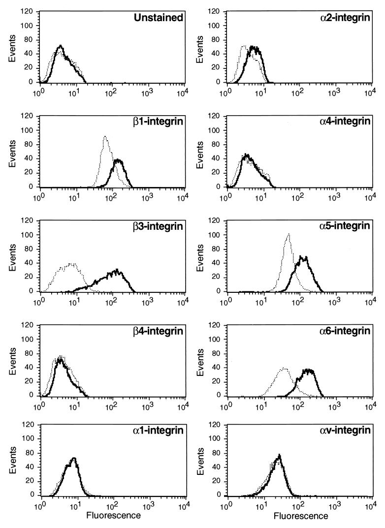
FIG. 2.
Expression of integrin subunits following Raf activation in NIH 3T3 cells. NIH 3T3 cells expressing ΔB-Raf:ER* were either untreated (dotted line) or treated with 100 nM 4-HT for 24 h (solid line) at which time the cells were harvested and either left unstained or stained for the cell surface expression of particular α- and β-integrin subunits as indicated. The expression of integrin subunits was detected by flow cytometry as described in Materials and Methods.

