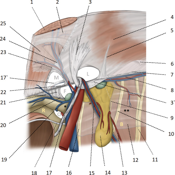
Fig. 1.
Anatomical basis of the myopectineal orifice (Fruchaud) or inner groin region. 1 Rectus abdominis; 2 Posterior rectus sheath with the arcuate line; 3 and 3’ Hesselbach’s ligament; 4 transverse muscle; 5 Henle’s ligament; 6 intermediate and endoabdominal fascia, respectively; 7 iliopubic tract; 8 A. and V. circumflexa iliaca interna; 9 genital branch of the genitofemoral nerve; 10 femoral branch genitofemoral nerve; 11 lateral femoral cutaneous nerve; 12 iliac fascia; 13 vascular supply of the lipoma from proximal and distal; 14 lipoma; 15 testicular vessels; 16 femoral nerve; 17 and 17’ deferent duct (in women the round ligament of uterus); 18 obliterated umbilical artery; 19 obturator vein; 20 pectineal ligament of Cooper; 21 lacunar ligament of Gimbernat; 22 the inner part of the inguinal ligament corresponds approximately to the iliopubic tract; 23 medial branch of the epigastric vessels, also known as corona mortis according to Hesselbach and Cooper; anastomoses between the retropubic vessels and the corona mortis are known as Bendavid’s circulus venosus; 24 inferior epigastric vessels; 25 fascia of the rectus abdominis. M medial hernia in Hesselbach’s triangle, L lateral hernia, internal inguinal ring, F femoral hernia, O foramen obturatorium, * triangle of doom (caveat: vascular injury), ** triangle of pain (caveat: nerve injury), R space of Retzius, B space of Bogros. Femoral and iliac lymph nodes are shown in green

