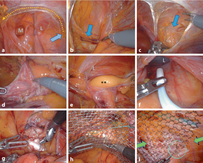
Fig. 4.
Surgical steps of robotic inguinal hernia repair (r-TAPP). a Opening of the peritoneum, starting laterally in projection of the anterior superior iliac spine (blue arrow), in a wing-like arc medially to the lateral umbilical fold. b Visualization of the pubic bone (blue arrow). c Visualization of the plane of the nerves under the iliac fascia, laterally. d Preparation of the hernia, in this case a lateral hernia, with monopolar dissection and separation of the hernia sac (*) from the deferent duct and the testicular vessels. e Exploration of the inguinal canal for the presence of lipoma (**) or preperitoneal fatty tissue. f In the case of long hernia sacs, the inguinal canal is sprayed with fibrin glue for seroma prophylaxis. g In the case of medial hernias, the transversalis fascia is reconstructed by suture, so that the posterior wall of the inguinal canal is flattened (caveat: do not damage the structures running behind the transversalis fascia). h Mesh fixation from medial to lateral, here at Cooper’s ligament. i Absorbable suture fixation of the mesh with a loose knot to the iliac fascia (needle in circle) under safe avoidance of the nerves (green arrows). M medial hernia/transversalis fascia, L lateral hernia/inner inguinal ring

