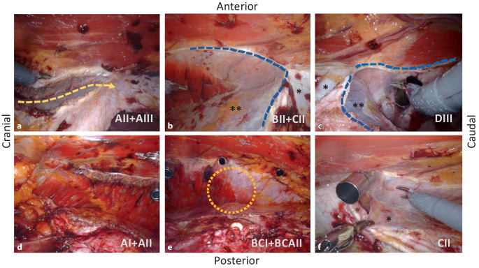
Fig. 3.
Intraoperative situs of robotic transversus abdominis release (r-TAR), left side of abdominal wall. The abbreviations in the right-bottom sections of the figure correspond to the grids of Fig. 1. a Release of the muscular insertion of the transversus abdominis in the area of the posterior rectus sheath (yellow dashed arrow). This corresponds to the entry from cranial or “top-down”. b Lateral detachment of the posterior rectus sheath (dashed blue line) and endoabdominal fascia/peritoneum from the transversus abdominis. c Entry to the transversus abdominis release from caudal or “down-to-up” (dashed blue line). d View after completion of the TAR in the region of the costal margin with the confluence of the diaphragm, the transversus abdominis and the rectus abdominis (see also Fig. 1). e View of the completed TAR on the left side; orange circle shows the demarcation of the fascia of the transversus abdominis, which remains cranial to the peritoneum and caudal to the muscle. f Detachment of the endoabdominal fascia and peritoneum from the transversus abdominis in the area of a port passage; this extraperitonealizes the port. *Posterior rectus sheath, **endoabdominal fascia with peritoneum

