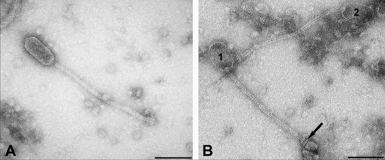
Figure 5.
Transmission electron micrograph of two distinct prolate phages resulting from Mitomycin C treatment of S. aureus CC1956 isolate WT19. A, Phage particle with pentagonal 38 nm in diameter capsid and a 12 nm thick tail with stacked disc appearance; B, Two phage particles (1, 2) with oval capsids of 55 nm in diameter and 9 nm thick tails with rail-road-track morphology. The base plate is separated from the tail by a transversal disc (arrow). Negative contrast preparation with uranyl acetate. Bars = 100 nm.

