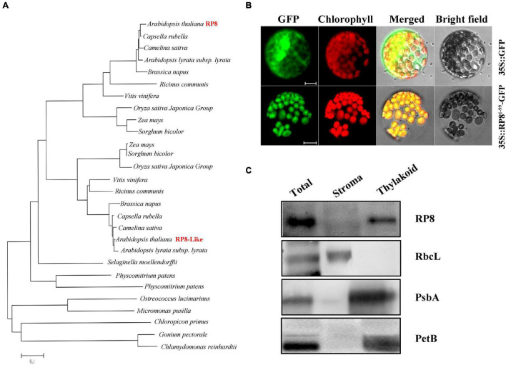
FIGURE 2.
Sequence, phylogenetic analysis, expression pattern, and subcellular localization of the RP8 protein. (A) Phylogenetic analysis of the RP8 protein and its homologs. Maximum likelihood analysis of the RP8 protein and its homologs from various organisms was performed. The unrooted phylogenetic tree was constructed by the Neighbor-Joining method (Saitou, 1987) with genetic distance calculated by MEGA 3.1. (B) Subcellular localization of RP8. Transit peptide of 99 amino acid residues at the N-terminus of the RP8 protein fused with GFP was transformed into N. benthamiana protoplasts by PEG transformation. The red-colored chlorophyll autofluorescence and GFP fluorescence were detected by a laser confocal-scanning microscopy after infiltration. Bars = 5 μm. (C) Immunoblot analysis of RP8 in chloroplast subfractions. Stromal and thylakoid proteins fractions were prepared and separated by SDS-PAGE. Immunoblot analysis using antibodies against RbcL, RP8, PsbA, and PetB was performed. Each lane was loaded with 30 μg proteins.

