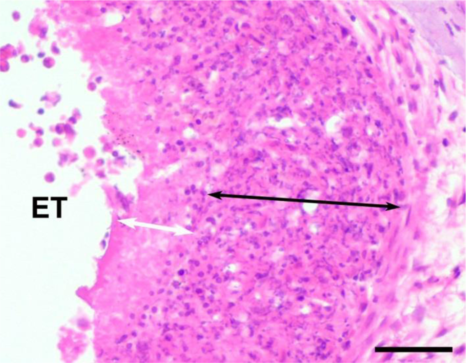
Figure 2.

Example illustrating the technique used to measure the width of the necrotic zone containing only cellular debris (white arrow), and the macrophage zone (black arrow). A necrotic zone was only occasionally observed, while a macrophage zone was commonly observed and located between the electrode array and loose fibrous tissue which typically occupied the remainder of the scala tympani. ET = electrode tract. Scale bar = 50 μm.
