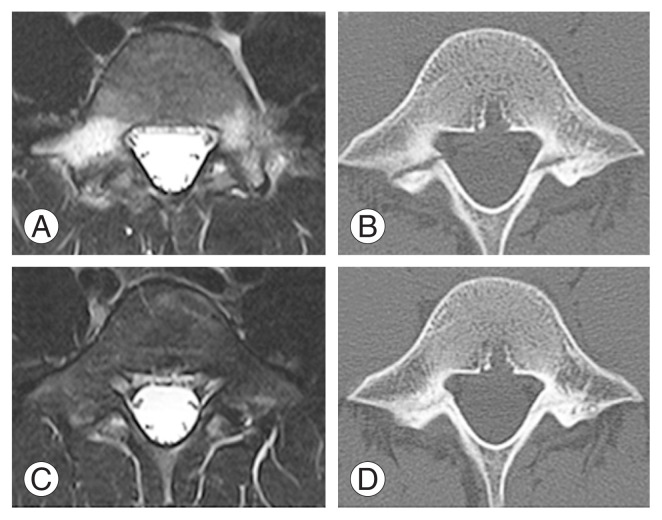
Fig. 3.
Fresh group: the case of a 14-year-old baseball player. (A) Magnetic resonance imaging revealed bilateral bone marrow edema at the first examination. (B) An axial computed tomography scan revealed a progressive-stage fracture on the right side and an early-stage fracture on the left side. (C, D) Three months later, we saw improved bone marrow edema and bone healing was achieved.

