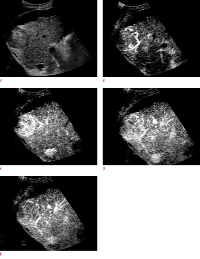
Fig. 5. A 67-year-old man with chronic liver disease and hepatocellular carcinoma.
A. B-mode ultrasonography indicates a hypoechoic nodule with well-defined limits. Note the presence of serrated ascites and liver contour. B-E. After contrast agent injection, contrast enhancement starts from the nodule periphery (B) and progresses centripetally (C) during the arterial phase. It is possible to observe a mild washout after 1 minute (D) during the portal venous phase. Mild and late washout (E) is compatible with the CEUS Liver Reporting and Data System 5 (CEUS-LI-RADS 5).

