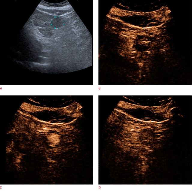
Fig. 6. A 53-year-old man with chronic liver disease and hepatocellular carcinoma.
A. B-mode ultrasonography shows a well-defined hypoechoic nodule in the chronic liver disease before contrast-enhanced ultrasonography (CEUS) evaluation. B, C. The arterial phase of CEUS shows nodule enhancement 20 seconds after contrast agent injection. D. In the portal phase, an enhanced nodule appears and, after 1 minute, shows mild washout (mild and late washout). This pattern is compatible with the CEUS Liver Reporting and Data System 5.

