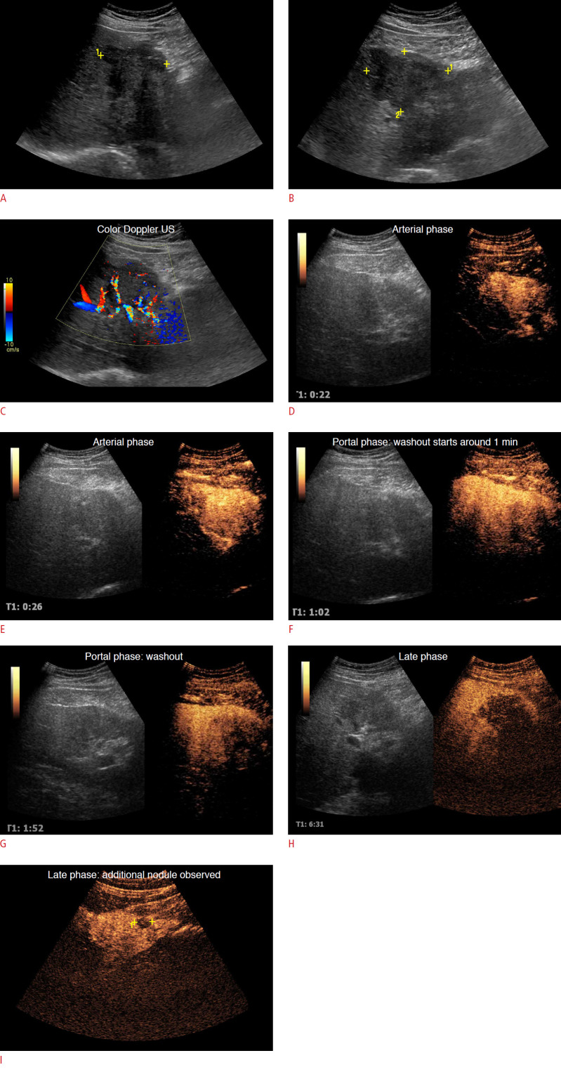
Fig. 8. A 56-year-old man, diagnosed with intrahepatic cholangiocellular carcinoma, peripheral subtype.
A, B. B-mode ultrasonography (US) shows an irregular and partially-defined hypoechoic nodule, measuring about 7.6×6.3×5.6 cm, located in segment IV. C. Color Doppler ultrasonography shows predominantly peripheral vascularization. D, E. Arterial phase: After contrast-enhanced ultrasonography, arterial enhancement is observed. F-H. Washout starts at 1 minute (F), and marked washout is observed in the portal phase (G), which becomes more pronounced in the late phases (H). I. It was possible to observe another satellite nodule (surrounded by calipers) in the late phase.

