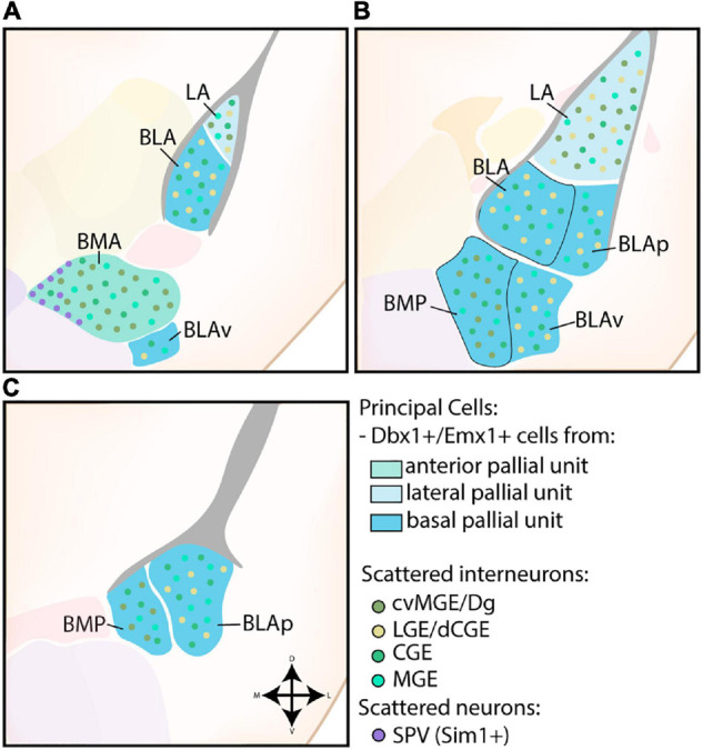
FIGURE 3.

Developmental origin of the basolateral complex. (A) The principal cells of the more anterior lateral amygdala (LA) and basolateral amygdala (BLA) are Dbx1-expressing neurons originating in the lateral and basal pallial units The anterior basomedial amygdala (BMA) contains a majority of Dbx1-expressing neurons from the anterior pallial unit, joined by Sim1-expressing neurons from the supraopto-paraventricular hypothalamus (SPV) at its medial side. In addition to these principal excitatory cells, some Emx1-expressing neurons are also present in the LA, BLA, and BMA at this level. The small part of the posterior ventral BLA (BLAv) contains a majority of excitatory Emx1-expressing cells from the basal pallial unit, with some Dbx1-expressing neurons scattered around (not shown). (B) The contributions from the Emx1-lineage increase along the rostrocaudal axis. At intermediate levels, more parts of the BLA become visible (BLAp and BLAv), both containing a majority of Emx1-expressing cells from the basal pallial unit, with some Dbx1-expressing cells still present (not shown). (C) At caudal levels, only the posterior subdivisions of the BM (BMP) and BLA (BLAp) are visible, both containing a majority of Emx1-expressing neurons with a minority of Dbx1-expressing neurons. Scattered interneurons can be found throughout the entire basolateral complex, with CGE and MGE-derived interneurons inhabiting the entire complex. In contrast, interneurons from the cvMGE/Dg area preferentially inhabit the LA and BM, while only data from the LA and BLA is available for dLGE/dCGE-derived interneurons from the Gsh2-lineage.
