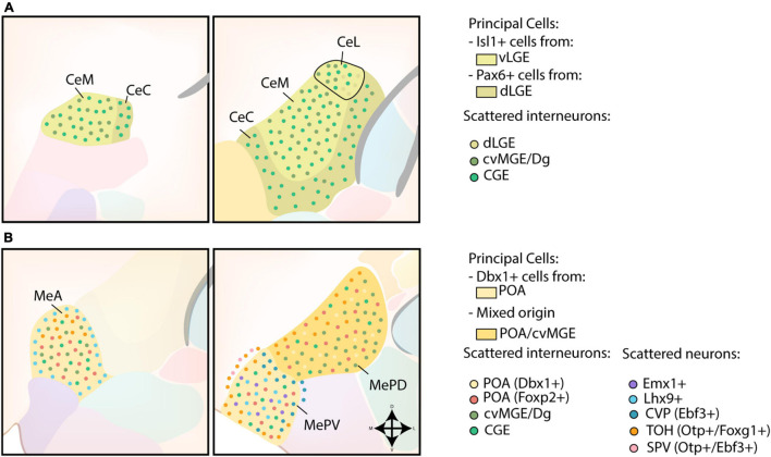
FIGURE 4.
Developmental origin of the centromedial amygdala. (A) Schematic overview of the neuronal origin of cells within the central amygdala (CA). Isl1-lineage cells from the ventral LGE (vLGE) inhabit the medial CA (CeM) and the centromedial part of the lateral CA (CeL), while Pax6-expressing cells from the dorsal LGE (dLGE) inhabit the capsular CA (CeC), with the exception of its ventromedial part, and the lateral part of the CeL. Presumptive CGE-derived interneurons can be found throughout the entire CA. Interneurons from the cvMGE instead prefer the CeM and CeL, and seem to avoid the CeC. (B) Schematic overview of the neuronal origin of cells within the medial amygdala (MA). POA-derived Dbx1-expressing neurons are spread throughout the entire medial amygdala (MA), but preferentially localize to the anterior MA (MeA) and posteroventral MA (MePV). They are complemented throughout the MA by Foxp2-expressing cells from the POA. Lhx9-expressing neurons, originating in the anterior amygdalar unit, populate a ring-shape surrounding the MeA and the core region of the MePV. Some Emx1-expressing cells were also identified in the MePV. Within the MePV, cells from the caudoventral pallium (CVP) and telencephalon-opto-hypothalamic domain (TOH) occupy specific niches. While cells from the TOH preferentially inhabit the medial side of an imaginary shell surrounding the MePV, cells from the CVP preferentially occupy the dorsolateral edge of the MePV. The TOH-derived cells are also present in the superficial layer of the MA, together with Otp+/Ebf3+ cells from other domains of the supraopto-paraventricular hypothalamus (SPV). TOH-derived neurons are also present within the posterodorsal MA (MePD), where they prefer to inhabit its medial part. A population of CGE-derived interneurons is presumed to populate the MA. In addition, small interneurons from the cvMGE/Dg are scattered within the MeA and the MePD.

