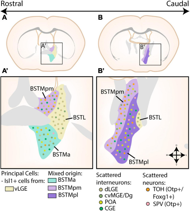
FIGURE 5.

Developmental origin of the Bed Nucleus of the Stria Terminalis. (A,A’) At rostral levels, the medial part of the bed nucleus of the stria terminalis (BSTM) contains a posteromedial (BSTMpm) and anterior (BSTMa) part. The entire BSTM contains inhibitory neurons derived from the caudoventral MGE or Diagonal area (cvMGE/Dg), CGE and preoptic area (POA). In addition, the BSTMpm contains a majority of telencephalon-opto-hypothalamic domain (TOH)-derived cells, although some cells originating from other supraopto-paraventricular (SPV) domains are also present. A small minority of TOH/SPV cells can also be found in the BSTMa. The lateral part of the bed nucleus of the stria terminalis (BSTL) is also visible at this level. The principal cells of this subnucleus are Isl1+ cells originating in the ventral LGE (vLGE), which are joined by some Pax6+ cells from the dLGE. Some of these Pax6+ cells were also found to invade the BSTM, while a minority of cvMGE-derived cells from the BSTM also inhabit the BSTL. (B,B’) At more caudal levels, the BSTM is divided into a posteromedial (BSTMpm) and posterolateral (BSTMpl) part. Here, the BSTMpm again contains the majority of TOH/SPV-derived cells, especially in its medial part, although the BSTMpl also contains scattered TOH/SPV-derived cells. The distribution of cvMGE/CGE/POA/dLGE/vLGE-derived cells in both the BSTM and BSTL remain similar to those at more rostral levels. Panels (A,B) are based on images from the Allen Brain Atlas, with the different subnuclei of the BST translated to extended amygdala terms via the scheme of Dong et al. (2001) and Allen Institute For Brain Science (2011).
