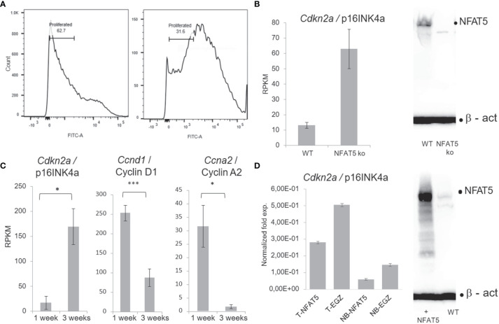
Figure 4.
NFAT5 supports the proliferation of KCs. (A) Measurement of KC proliferation cultivated for one week (left) or three weeks (right) in CFSE assays. (B) Increase of p16INK4a RNA levels in KCs upon NFAT5 ablation. The western blot (right) confirms the absence of NFAT5 expression in KCs isolated from Nfat5-/- mice. (C) Expression of cell cycle inhibitor p16INK4a and of cyclins D1 and A2 in KCs cultivated for one or three weeks. Next-generation-sequencing (NGS) data of four independent batches of KCs cultured for 1 or 3 weeks are shown. (D) Decrease in p16INK4a RNA levels in KCs isolated from the tails of adult (T) or from newborn mice (NB). The western blot illustrates increased NFAT5 levels in tail KCs that have been transduced with a retrovirus expressing NFAT5. The KCs in (C, D) were cultivated for 1 week. (***p<0.0001, **p<0.001 and *p<0.05).

