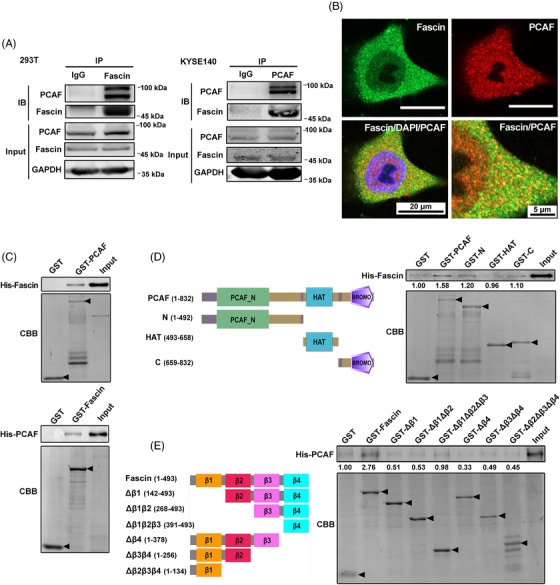
FIGURE 1.

Fascin interacts with PCAF in ESCC cells. A, The interaction between Fascin and PCAF was investigated by immunoprecipitation assay using anti‐Fascin and anti‐PCAF antibodies in HEK293T and KYSE140 cells, respectively. B, Immunofluorescence analysis of the localization of endogenous Fascin and PCAF in ESCC KYSE150 cells. Endogenous Fascin (green) and PCAF (red) were stained with specific antibodies, and nuclei were stained with DAPI (blue). C, Fascin directly interacted with PCAF in in vitro GST‐pull down assays using GST‐PCAF and GST‐Fascin. D, The N‐terminal domain, acyltransferase domain (HAT), and C‐terminal BROMO domains of PCAF were fused to GST and purified for GST‐pull down assays with His‐fused Fascin (His‐Fascin). E, The GST‐fused truncated forms of Fascin were purified for GST‐pull down assays with His‐fused PCAF (His‐PCAF). The purified GST fusion proteins were examined with Coomassie brilliant blue staining, and the pulled‐down His proteins were examined by Western blotting. Arrows indicate proteins with correct molecular weights. Abbreviations: PCAF: P300/CBP‐associated factor; ESCC: esophageal squamous cell carcinoma; IP: immunoprecipitation; IB: immunoblot; GAPDH: glyceraldehyde‐3‐phosphate dehydrogenase; DAPI: 4’,6‐diamidino‐2‐phenylindole; CBB: Coomassie brilliant blue; GST: Glutathione S‐transferase; HAT: Histone acyltransferase; BROMO: bromodomain
