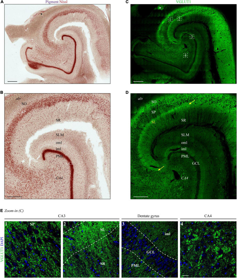
FIGURE 1.
Layers of the human hippocampus determined by VGLUT1 immunofluorescence and Nissl staining. (A) Lipofuscin (pigment, purple) and Nissl substance (red) were stained with aldehyde fuchsine and Darrow red in a human hippocampal section, occipital part. The asterisk marks adjoining parts of the gyrus fasciolaris. Scale bar, 700 μm. (B) Zoom-in of panel (A) to display layer boundaries. Alv = alveus, SO = stratum oriens, SP = stratum pyramidale, SR = stratum radiatum, SLM = stratum lacunosum-moleculare, oml = outer molecular layer, iml = inner molecular layer, PML = polymorphic layer, GCL = granule cell layer, CA4 = cornu Ammonis region 4. Scale bar, 500 μm. (C) A subsequent section from the tissue block used in panel (A) was immunostained with VGLUT1 and acquired at a confocal microscope. LAS-X Navigator was used to create and stitch the tilescan. Dashed squares (white) are shown in panel (E) as high-resolution z-stacks. The asterisk marks adjoining parts of the gyrus fasciolaris. Scale bar, 700 μm. (D) Zoom-in of panel (C) to display layer boundaries. The yellow arrows mark the borders of the stratum lucidum (SL), i.e., CA3. Scale bar, 500 μm. (E) Maximum intensity projections (MIPs) of high-resolution z-stacks (stack size = 2.64 μm) acquired in the SP of CA3 (1), SL and SR of CA3 (2), dentate gyrus (3), and CA4 (4). Numbers refer to position 1–4 in the tilescan from panel (C). Scale bar, 30 μm. Images in panels (A–E) are derived from case 1.

