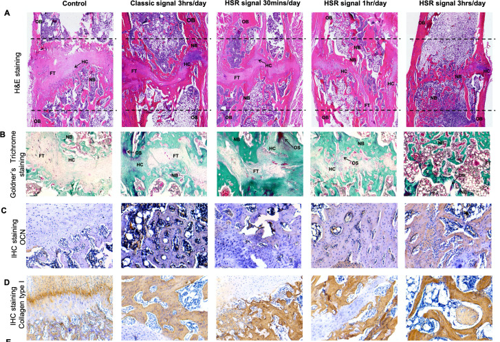
Fig. 9.
Histological and immunohistochemical results of the distraction site on week 6 after distraction. a) Haematoxylin and eosin (H&E) staining of the distraction site.Each figure was obtained by stitching 20 adjacent images of the distraction site (50× magnification). b) Goldner’s Trichrome staining. Magnification: 50×. c) Immunohistochemical (IHC) staining of osteocalcin (OCN). Magnification: 100×. d) IHC staining of collagen type I (Col I). three one FT, fibrous tissue; HC, hyaline cartilage; HSR, high slew rate; NB, new bone; OB, old bone; OS, osteoid.

