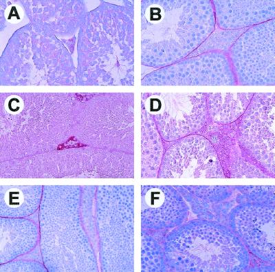
FIG. 4.
Impaired differentiation of Leydig cells from Ink4c-null and Ink4cd double-null mice. P450scc staining of Leydig cells is indicated by red precipitate. (A) Immature Leydig cells from 1-month-old wild-type mice lack P450scc staining. (B) Absence of staining of 3-month-old wild-type type testis with preimmune serum used as negative control. P450scc staining in Leydig cells from 3-month-old wild-type (C) and Ink4d-null (D) mice. Testes from 3-month-old Ink4c-null (E) and Ink4cd doubly deficient (F) mice failed to reveal significant P450scc staining.

