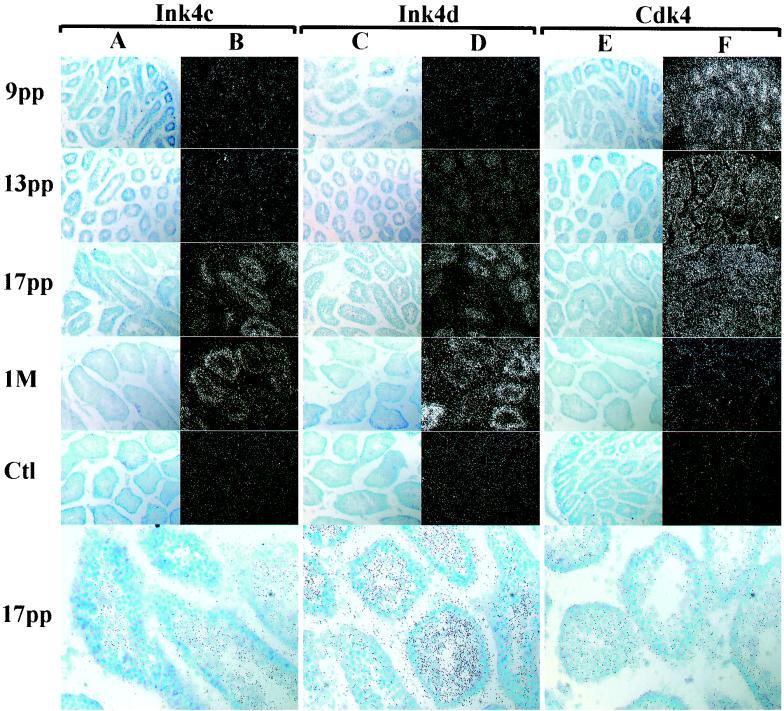
FIG. 6.
Ink4c, Ink4d, and Cdk4 mRNAs expressed during normal testis development. In situ hybridizations were performed on sections of testes taken from wild-type animals at 9, 13, and 17 days and 1 month (M) pp as indicated at the left of the panels. Sections were hybridized with the antisense probes indicated at the top. Morphology of the tubules is revealed in bright-field images (A, C, and E), and the hybridization intensity is revealed by dark-field images (B, D, and F). Control hybridizations (Ct1) performed with sense strand probes at 1 month pp (Ink4c and Ink4d) and at day 9 pp (Cdk4) are shown. All magnifications are ×200. The three bottom bright-field panels indicate the localization of grains within tubules, as photographed at higher power (magnification, ×400).

