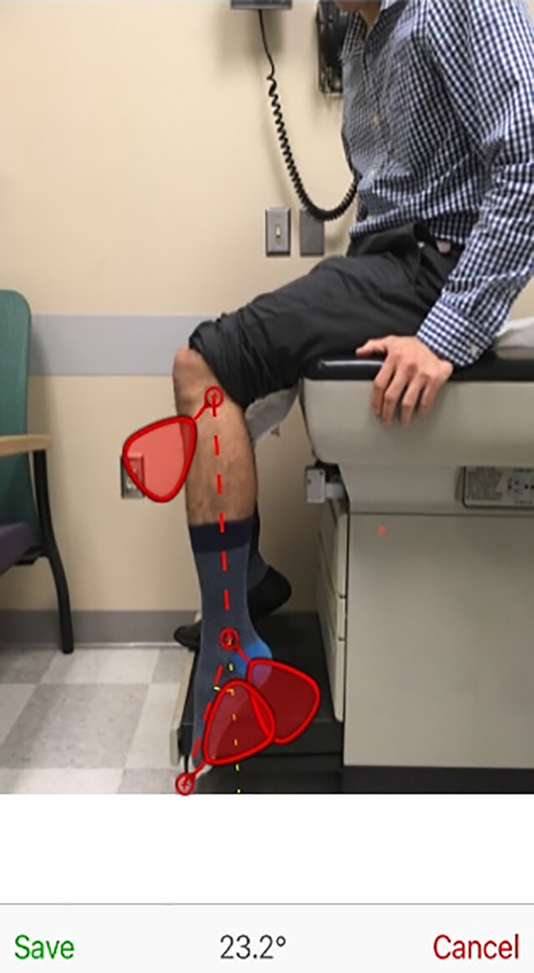
Figure 6.

For the DrG measurements, after a picture was taken with the participant’s leg, foot, and ankle in the frame, the examiner placed the 3 movable red markers on the participant’s fibular head, lateral malleolus, and just above the fifth toe such that the line between the markers of the lateral malleolus and fifth toe was parallel to the longitudinal axis of the participant’s fifth metatarsal. The angle of plantarflexion or dorsiflexion was documented as displayed on the bottom of the screen after marker placement.
