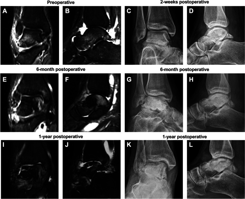
Figure 1.
Coronal and sagittal magnetic resonance images of a 25-year-old woman before (A, B) and 6 months and 1 year after undergoing a talar subchondroplasty and open Broström procedure 2 weeks after the date of injury. Corresponding anteroposterior and lateral radiographs are shown at time points of (C, D) 2 weeks, (E-H) 6 months, and (I-L) 1 year after the same procedure. Postoperatively there is evolution from talar bone marrow edema and sclerosis to collapse and fragmentation.

