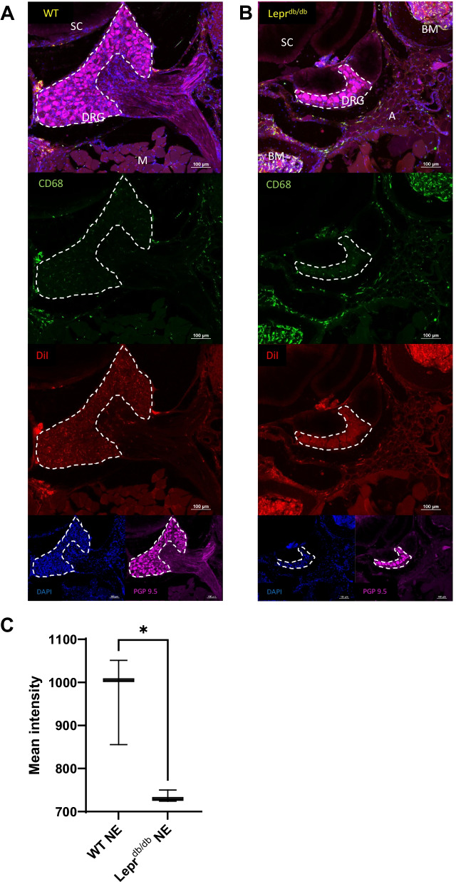
Fig. 11.
Identification CD68+ and DiI+ cells within the lumbar DRGs of WT and Leprdb/db mice. Sections containing lumbar DRG tissue from WT (A) and Leprdb/db mice (B) were reviewed to determine the location of CD68+DiI+ cells. CD68+DiI+ double positive cells were identified within the DRG, perineural adipose tissue, and meninges surrounding the DRGs. Analysis of the DiI mean signal intensity within the DRG itself revealed a significant difference between WT (n = 3; C) and Leprdb/db (n = 3; D) mice (p = 0.0167). Images were taken at 20 × and a composite image was formed using the tile scan feature of the Nikon confocal microscope. Scale bar = 100 µm. Error bar represents the standard deviation from three independent measurements

