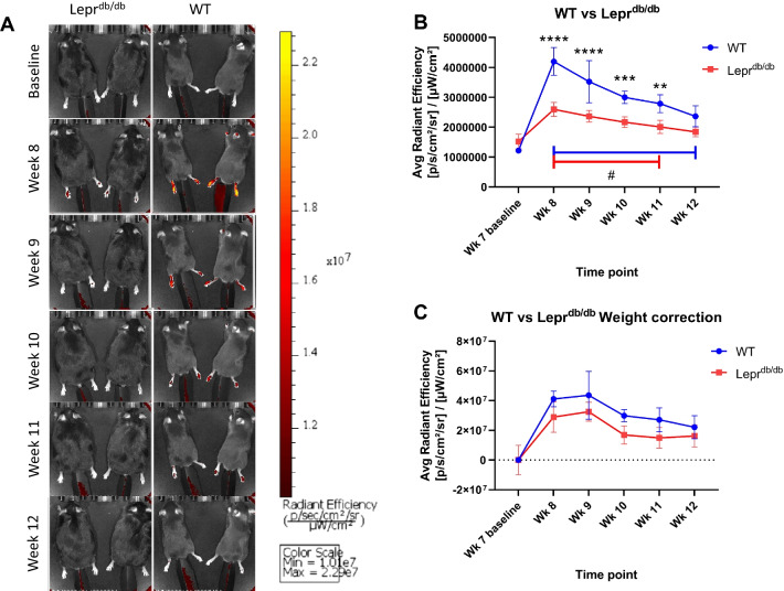
Fig. 4.
DiR signal is detectable in vivo using IVIS imaging. WT (n = 5) and Leprdb/db (n = 5) mice were injected i.v. with 200 µl of PFC-NE. Images were taken using an IVIS Lumina system from 7 weeks of age (baseline) until 12 weeks of age (A). Injections were performed 72 h prior to the 8 week images. Fluorescence from the DiR component of the PFC-NE was detected within foot pads of each mouse and was tracked over time as Avg Radiant Efficiency ([p/s/cm2/sr]/[µW/cm2]) (B). A weight correction was performed to determine the extent to which differences seen between WT and Leprdb/db mice were caused by differences in body mass (C). **(p < 0.01), ***(p < 0.001), ****(p < 0.0001). Error bar represents the standard deviation from five independent measurements

