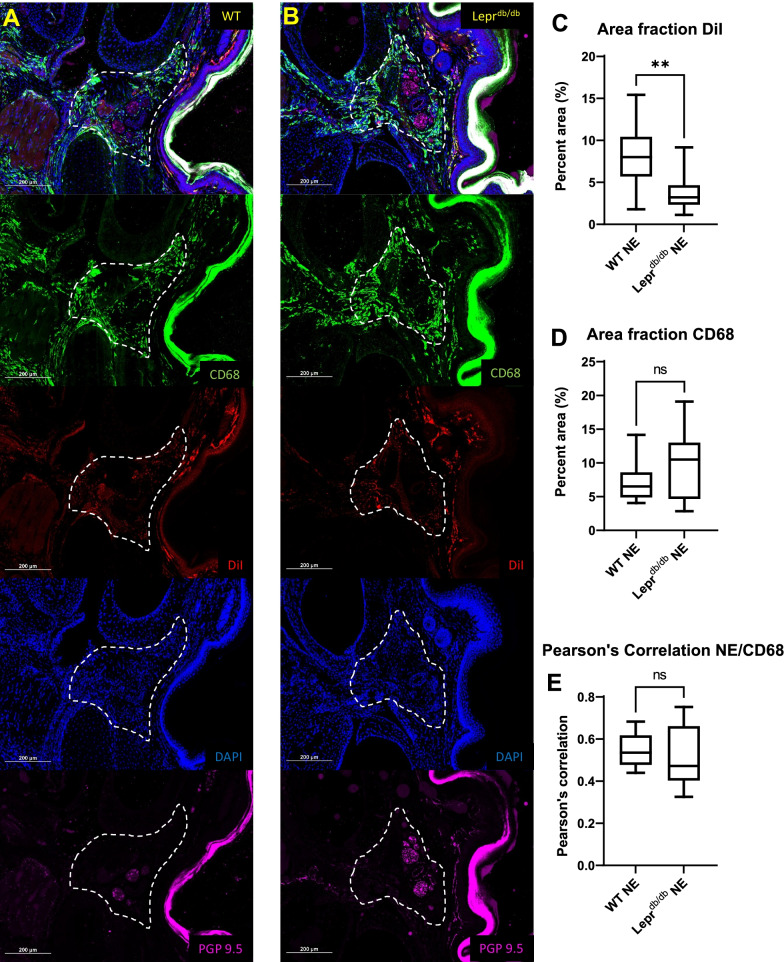
Fig. 7.
Quantification of CD68 and DiI signal in hypodermal tissue surrounding the Neurovascular bundles of the hind limb foot pad. Analysis of the hypodermal regions (dotted white line) between metatarsals 2, 3 and 4 from hind limb foot pads of WT (n = 3) (A) and Leprdb/db (n = 3) (B) mice showed a significantly lower amount of DiI surrounding the nerves (N) of neurovascular bundles of Leprdb/db mice as compared to WT mice (p = 0.0063) (C). No significant difference was found for percent area of CD68 (p = 0.3389) (D) or Pearson’s correlation for CD68 and DiI (p = 0.7317) (E) when comparing WT and Leprdb/db mice. Scale bar = 200 µm. Error bar represents the standard deviation from twelve independent measurements

