
Fig. 1.
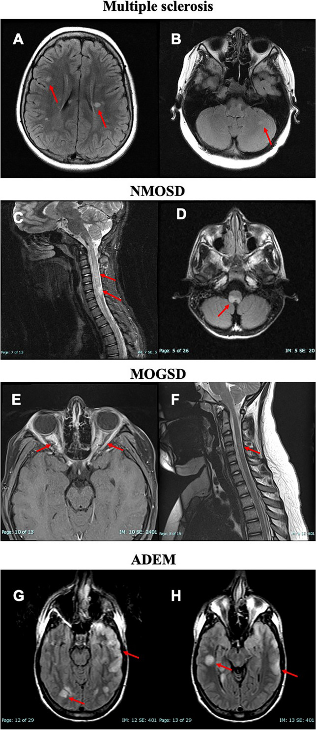
MRI-based differential diagnosis of pediatric patients with multiple sclerosis, neuromyelitis optica spectrum disorder, myelin oligodendrocyte spectrum disorder, and acute demyelinating encephalomyelitis. A, B Axial FLAIR images demonstrating typical MS-based hyperintensities within the periventricular region, juxtacortical region, and in the posterior fossa (cerebellum). C, D Sagittal T2 image of the spinal cord and axial FLAIR image demonstrating longitudinal extensive transverse myelitis that involves the brainstem region and extend over the entire cervical portion of the spinal cord. E, F Axial post-contrast FLAIR image demonstrating bilateral optic neuritis with optic nerve edema associated with contrast enhancement of the optic nerve and perineural sheet. Coronal T2 image demonstrating cervical spinal cord lesion spanning over 2.5 vertebral levels. G, H Axial FLAIR images demonstrate multiple, diffuse, ‘fluffy,’ poorly defined T2 hyperintense lesions involving both grey and white matter and both supra- and infratentorial region. ADEM acute disseminated encephalomyelitis, FLAIR fluid-attenuated inversion recovery MOGSD myelin oligodendrocyte glycoprotein antibody spectrum disorder, MS multiple sclerosis, NMOSD neuromyelitis optica spectrum disorders