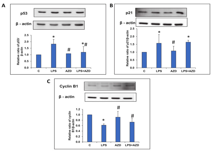
Figure 6.
LPS-induced alterations in the expression of cell cycle regulatory markers. Protein extracts from Rin-5F cells treated with LPS with/without AZD were separated on 12% SDS-PAGE and transferred on to nitrocellulose paper by Western blotting. Transferred proteins were detected using specific antibodies against p53 (A), p21 (B), and cyclin B1 (C), and visualized by enhanced chemiluminescence using the Sapphire Biomolecular Imager (Azure biosystems, Dublin USA) or using X-ray films. Beta actin was used as loading control. Histograms represent the relative ratios of the quantitated proteins normalized against the loading control. The figures are representative of at least three individual repetitive experiments. Asterisks indicate significant differences fixed at p ≤ 0.05 (* indicates significant difference relative to control untreated cells, whereas # indicates significant difference relative to LPS-treated cells).

