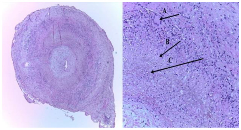
Figure 1.
The histopathologic picture of the left superficial temporal artery biopsy (TAB): (A) intimal thickening, and an inflammatory infiltrate with giant cells of the media layer (typical granulomatous inflammation), (B) epithelioid cells, and (C) characteristic internal limiting lamina fragmentation (H&E staining-left-×40; right-×100) [9].

