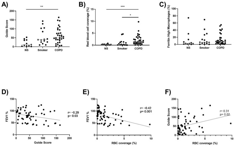
Figure 1.
Lung macrophage iron levels and RBC coverage is increased in COPD. (A) Lung tissue sections were stained with Perls Prussian Blue. Lung macrophage iron levels were quantified in 11 NS, 15 S and 32 COPD patients. (B) H&E staining was carried out to assess RBC coverage in the alveolar space in 11 NS, 15 S and 32 COPD patients. (C) The percentage of ferritinhigh lung macrophages in 11 NS, 15 S and 32 COPD patients. (D) Correlation between FEV1% and Golde score (r = −0.29, p = 0.03). (E) Correlation between FEV1% and RBC coverage of the alveolar space (r = −0.42, p = 0.001). (F) Correlation between Golde score and RBC coverage of the alveolar space (r = 0.31, p = 0.02). Kruskal–Wallis multiple comparisons test was used to test differences between groups. Correlation data are plotted as individuals with linear regression (D–F). * = p < 0.05, ** = p < 0.01, *** = p < 0.001. Data presented as individuals with median.

