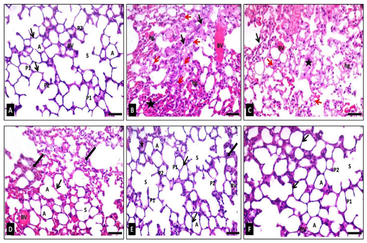
Figure 8.
Photomicrographs of sections of the lung tissues of adult male mice stained with H&E showing: (A) Group I (control): alveolar sacs (s), alveoli with thin walls (arrows) formed of flat cells with densely stained nuclei type I pneumocytes (P1) and type II pneumocytes (P2) with large rounded nuclei, clear alveolar spaces (A), small thin-walled blood vessels (BV). (B) Group II (diseased): the lung architecture is disordered; some alveoli appear collapsed (star) with very thick alveolar walls (black arrows). Many alveolar epithelial lining cells show vacuolar cytoplasmic necrosis with pyknotic nuclei (red arrows). Severely congested blood vessels (BV) and interstitial hemorrhages (hg) could be seen. (C) Group III (diseased, 50 mg/kg FEE): moderate disorganization of alveoli; collapsed alveoli (star) with moderate thickening of the alveolar wall (black arrow) and many epithelial linings of the alveoli with pyknotic nuclei and vacuolated cytoplasm (red arrows). Moderately congested blood vessels (BV) and interstitial hemorrhages (hg) could be observed. (D) Group IV (diseased, 100 mg/kg FEE): some normal alveolar sacs (S) and alveoli (A) with thin walls (thin arrows). Notice, mild thickening of alveolar walls (thick arrows), congested blood vessels (BV), and minimal interstitial hemorrhages (hg). (E) Group V (diseased, 150 mg/kg FEE): the lung architecture is relatively near to the normal with alveolar sacs (S), clear alveolar spaces (A), thin-walled alveoli (thin arrows), normal type I pneumocytes (P1), as well as type II pneumocytes (P2) and small thin-walled blood vessels (BV). Few alveoli are collapsed (star) with minimal thick walls (thick arrow). (F) Group VI (150 mg/kg FEE only): the architecture of the lung is well organized. The alveolar sacs (S), alveolar spaces (A), the alveolar walls (arrows), type I pneumocytes (P1) and type II pneumocytes (P2) and blood vessels (BV) appear normal. [H&E ×400, scale bar = 50 μm].

