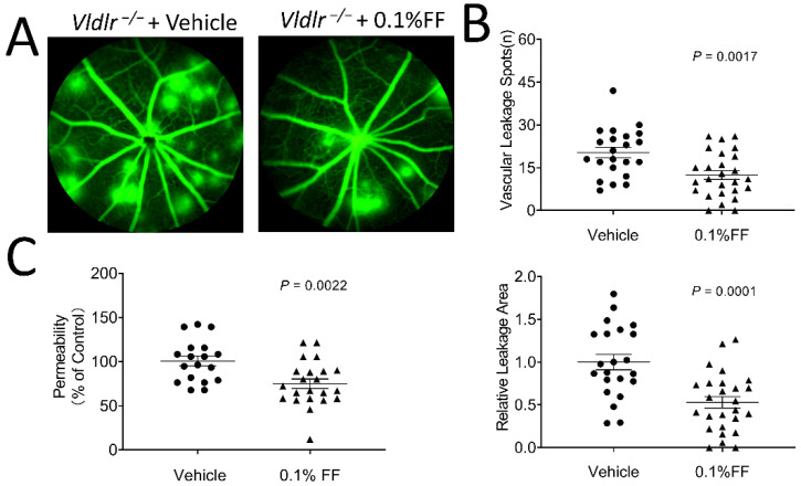
Figure 2.
Fenofibrate eye drops alleviate fundus vascular leakage and permeability in Vldlr−/− mice. The animals received 0.1% fenofibrate (FF) eye drop treatment (twice a day) for 7 days. Fundus fluorescein angiography was performed, and retinal vascular permeability was quantitated using Evans blue as a tracer. (A) Representative images showed the fundus vascular leakage (hyperfluorescent spots) corresponding to the areas of increased choroidal and retinal permeability. (B) The vascular leakage was quantified by the number of hyperfluorescent spots and the percentage of hyperfluorescent area in total retinal area in the 0.1% FF group (n = 26 eyes in each group) and the vehicle group (n = 22 eyes in each group). (C) The permeability of the retinal vasculature was measured in the 0.1% FF eye drop group (n = 22 eyes in each group) and vehicle group (n = 18 eyes in each group) using Evans blue dye as a tracer. The extracted Evans blue dye was normalized to the total retina protein. Data were expressed as mean ± SEM, analyzed by an unpaired Student’s t test.

