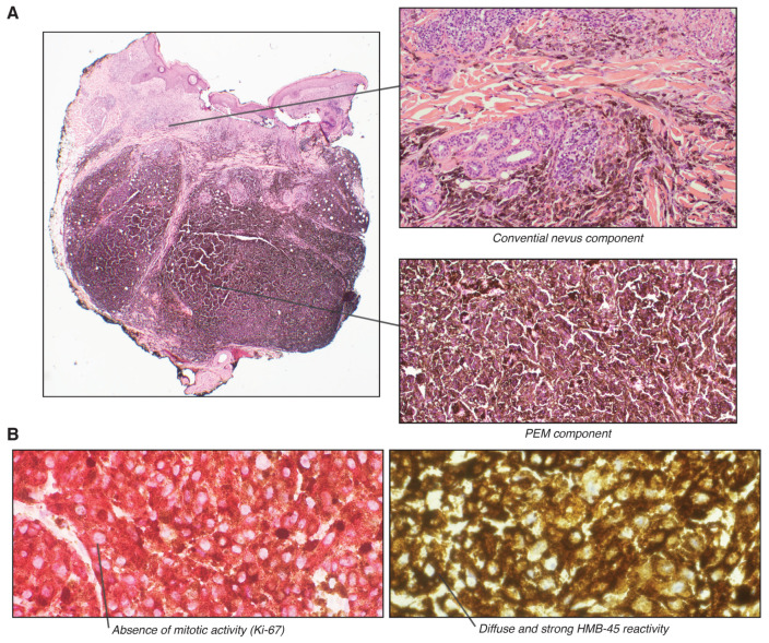
Figure 2.
Microscopic examination. (A). (left) Low-power view of the tumor, presenting as a 4 mm nodule located in the deep dermis and extending into the subcutaneous fat. (top right) Heavily pigmented melanocytes have epithelioid and spindled appearance. (bottom right) Combined blue nevus-like component. (B). (left) Ki-67 immunohistochemistry showing absence of mitotic activity. (right) HMB-45 immunohistochemistry shows diffuse and strong reactivity in tumor cells.

